Image 3
|
Image 3 |
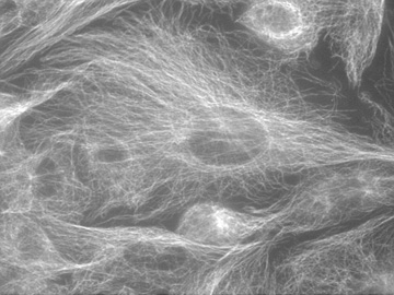
| Image of L8 cells growing on a coverslip. The cells were fixed, permeabilized, and then labeled with a mouse primary antibody to tubulin, and subsequently with an anti-mouse second antibody conjugated to the fluorochrome fluorescein. The cells are imaged using fluorescent illumination employing a fluorescein filter cube. When viewed in the microscope the white objects appeared green; however the picture is black and white. The microtubule arrays in the cells are clearly visible. The microtubules run from the cell membrane to organizing centers (microtubule organizing centers) located near the cell nucleus. Since the nuclei contain no microtubules, they appear as hollow structures inside the cells. |
| Click on the image to return to the previous page. |