Images 4
|
 |
|
Image 4a |
Image 4b | |
 |
 |
|
| Image 4c | Image 4d | |
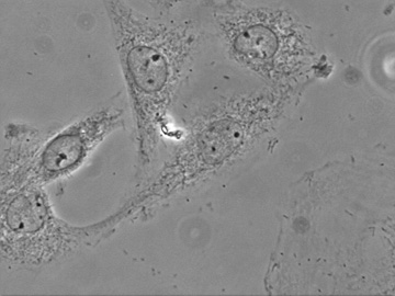
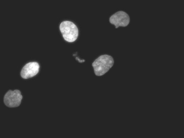
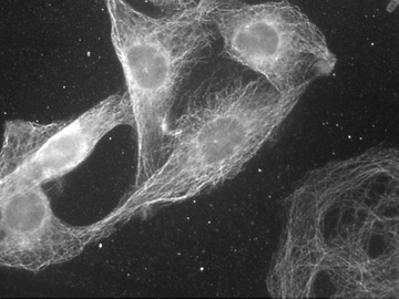
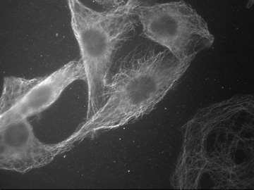
| Images of L8 cells growing on a coverslip. The cells were fixed, permeabilized, and then labeled with a series of reagents: DAPI (a nuclear stain), a mouse antibody to tubulin and subsequently an anti-mouse second antibody conjugated to fluorescein, a rabbit antibody to an intermediate filament protein known as vimentin and subsequently an anti-rabbit antibody conjugated to rhodamine. The first image (4a) is a picture of the cells seen in phase contrast. The nuclei, nucleoli, and structures within the cytoplasm are visible. Note how the cells appear rather fusiform (football-shaped) and spread out on the surface of the coverslip. For the second and subsequent images, the transillumination was turned off and the fluorescent illumination turned on. The second image (4b) is a picture of the same field viewed through the DAPI filter cube. The stained objects (the nuclei) appeared blue in the microscope. The third image (4c) is a picture of the field viewed through the fluorescein filter cube. The microtubules in the cells are visible. These tubules appeared green in the microscope. Each bright spot over the nucleus in two of the cells near the center represents a microtubule organizing center. The fourth image (4d) is a picture of the field viewed through the rhodamine filter cube. Seen here are the intermediate filaments connecting the cell membrane and nucleus. The filaments appeared red in the microscope. By switching back and forth between the various filter cubes, all these objects could be viewed. A colored picture representing the superimposition of the DAPI, fluorescein, and rhodamine images is presented as image 5. | ||
| Click on any image to return to the previous page. | ||