Image 5
|
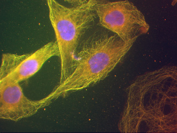
Image 5 |
| This image should be viewed in color. It represents the superposition of each of the images 4b, 4c, and 4d. Each individual image was first colored to correspond to the color seen in the microscope. The colored images were then combined into one image. Thus, blue represents the DAPI-stained nuclei, green represents the antibody-stained tubulin, and red represents the antibody-stained vimentin. For further explanation see the legend to the items in Images 4b-d. |
| Click on the image to return to the previous page. |