Images 7 & 8
 |
 |
|
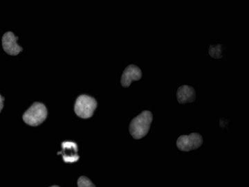
Image 7 |
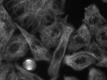
Image 8 |
|
| These two images are of L8 cells. Image 7 shows the DAPI label (this appeared blue in the microscope). Note that most of the cells have rather uniformly labeled nuclei; these cells are in interphase. However, the cell toward the bottom left is in mitosis (probably late anaphase). In the microscope, individual chromatids were much more visible. Image 8 shows the same field and same cells labeled for tubulin (for details see the text accompaning Image 3). Most of the cells here are out of focus. However the cell undergoing mitosis is in focus. Note the spindle apparatus that is visible in this cell. Note also that the cell has rounded up in preparation for mitosis; the remaining cells are flattened against the coverslip and appear fusiform. Image 9 is a colored superposition of these two images. | ||
| Click on any image to return to the previous page. | ||