Images 1 & 2
 |
 |
|
Image 1 |
Image 2 |
|
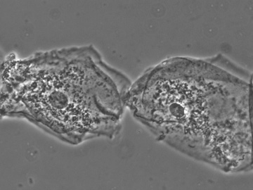
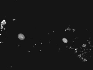
| These two images were obtained of human cheek epithelial cells. Image 1 is obtained by using phase contrast. The nucleus in each cell and the cell membranes are visible. Image 2 is obtained from the same field as Image 1. The illumination from light source in the base of the microscope was turned off and the fluorescent illumination (using the filter cube for rhodamine) was turned on. Since the dye propidium iodide (which stains nucleic acids) was applied to these cells, the nuclei and bacteria on the surface of the cells are visible. When viewed through the microscope the objects seen in fluorescence were red, but appear white on black here because the camera was black and white. | ||
| Click on any image to return to the previous page. | ||